

| Host / Isotype | Clonality | Applications | MW (kDa) | Reactivity |
| Rabbit IgG | Polyclonal | WB, IHC-P, IF/ICC, ELISA | 42 | Human, Mouse, Rat, Monkey, Pig, Chicken, Sheep, Hamster |
| Application Key | WB-Western IP-Immunoprecipitation IHC-Immunohistochemistry IF-Immunofluorescence ICC-Immunocytochemistry FC-Flow Cytometry |
| Reactivity Key | H-Human M-Mouse R-Rat Mk-Monkey B-Bovine Pg-Pig Hm-Hamster Dg-Dog C-Chicken X-Xenopus Z-Zebrafish Hr-Horse All-All Species Expected |
| Applications Dilutions | |
| The application notes include recommended starting dilutions;optimal dilutions/concentrations should be detemined by the end user. | |
| WB(Westem Blotting) IHC-P(Immunohistochemistry-Paraffin) IF/ICC(Immunofluorescence/Immunocytochemistry) ELISA(Enzyme-linked Immunosorbent Assay) |
WB=1:1000-5000 IHC-P=1:50-300 IF/ICC=1:50-300 |
| Product Information | |
| Conjugate | Unconjugated |
| Specificity | β-Actin Rabbit Polyclonal Antibody detects endogenous levels of β-Actin protein. |
| Purification | Affinity purification |
| Concentration | 1mg/ml |
| Format | Liquid |
| Formulation | In PBS, pH 7.4, containing 0.02% sodium azide,0.5% BSA and 50% glycerol. |
| Shipping | Gel Pack |
| Storage | Store at -20°C least 1 year from the date of shipment. avoid repeated freeze/thaw cycles. Aliquots may be stored at +4°C for 1-2 weeks. |
| UniProt ID | |
| Entrez Gene ID | |
| Product Description | |
β-Actin is a principal component of the cytoskeleton, forming microfilaments that are essential for maintaining cell shape, enabling cell movement, and facilitating intracellular transport. β-Actin exists in a dynamic equilibrium between monomeric globular actin (G-actin) and filamentous actin (F-actin), allowing for rapid reorganization of the cytoskeleton in response to cellular signals. This reorganization is crucial for processes such as muscle contraction, cell division, and cell signaling. The regulation of β-Actin polymerization and depolymerization is controlled by small GTPase family members like Rho, Rac, and Cdc42, which influence the formation of actin stress fibers, membrane ruffles, and filopodia, respectively. These processes are vital for cell adhesion, migration, and morphology. |
| Product Image Gallery | |
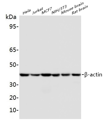 Western blot analysis of extracts on Hela, Jurkat, MCF7, NIH/3T3, Mouse brain, Rat brain cels, using β-Actin Rabbit Polyclonal Antibody (EAB10002) at 1:2000 dilution. Secondary antibody Goat Anti-Rabbit IgG (H&L)-HRP (EAB21002) at 1:5000 dilution.
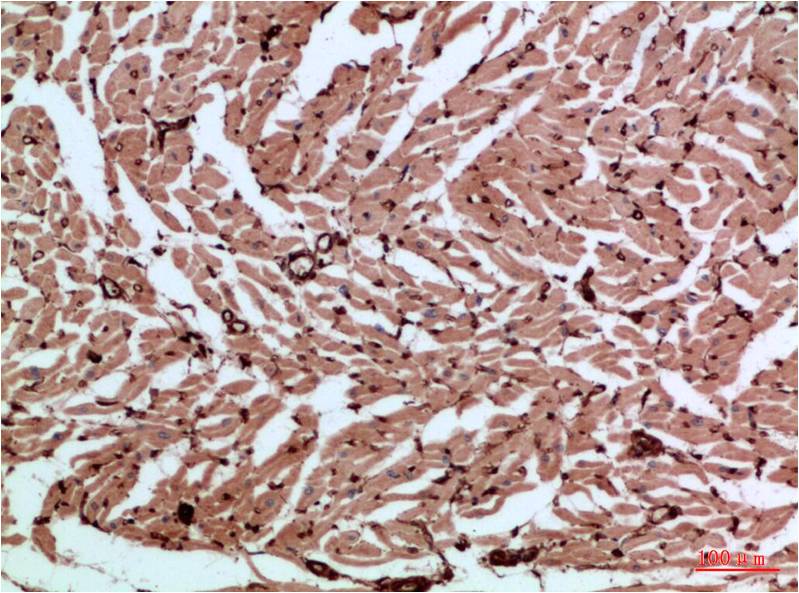 Immunohistochemical analysis of paraffin-embedded Human heart tissue, using β-Actin Rabbit Polyclonal Antibody (EAB10002) at 1:200 dilution.
|
